IHC Biomarker Scoring
Quantify HER2, PD-L1, and Ki-67 in 90 Seconds
Synthia analyzes whole-slide biopsy images automatically — delivering reproducible IHC scores before the next case is staged.
The Problem
Manual IHC Scoring Is the Diagnostic Bottleneck
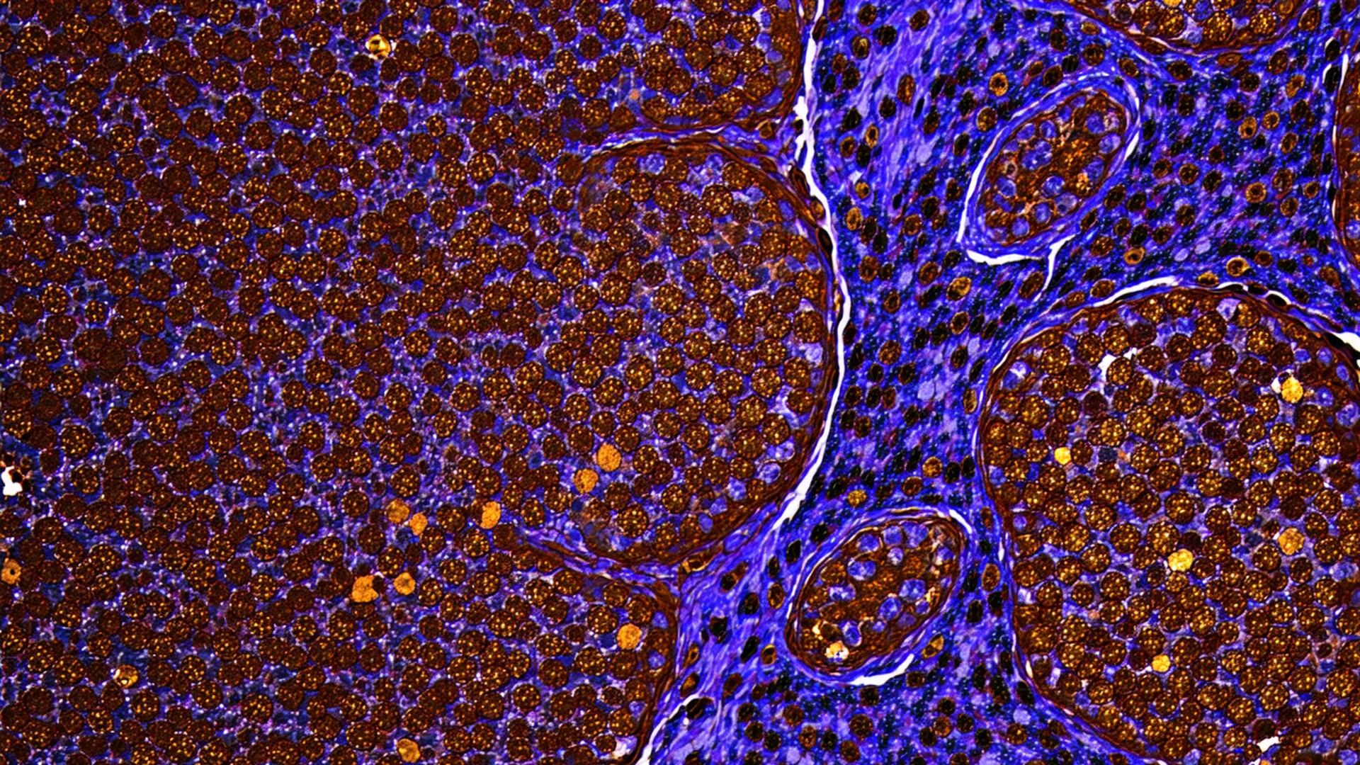
Immunohistochemistry scoring is essential for HER2, PD-L1, and Ki-67 treatment eligibility decisions — yet it remains one of the most subjective, time-intensive steps in the oncology diagnostic workflow. A single breast cancer biopsy batch can require 30–90 minutes of manual annotation per pathologist.
The consequences are real: inter-pathologist variability of 15–25% on HER2 equivocal cases, delayed treatment decisions, and burned pathologist time that should be spent on diagnostic interpretation — not counting brown cells.
Synthia was built for one purpose: automate the quantification so pathologists can focus on the judgment.
How Our Model WorksIHC DIAGNOSTIC WORKFLOW
Process
How Synthia Works
Three steps from WSI upload to structured biomarker report.
Upload WSI
Drag-and-drop NDPI, SVS, or TIFF whole-slide images — or connect via your PACS or LIS. Synthia accepts the major scanner formats from Hamamatsu, Leica/Aperio, and 3DHISTECH.
AI Scores Tissue
Synthia's deep learning model localizes IHC-positive cells, quantifies DAB staining intensity, and calculates a standardized score per ASCO/CAP guidelines — automatically, without human annotation.
Pathologist Reviews Report
Receive a structured PDF and structured data report in under 90 seconds. Review the quantified scores, annotate any disagreements, and sign off in one click. You interpret. We quantify.
Three Biomarkers. One Workflow.
Each biomarker scored to its clinical standard — HER2 per ASCO/CAP, PD-L1 per assay-specific thresholds, Ki-67 with hotspot detection.
Breast & Gastric Cancer
HER2
0 / 1+ / 2+ / 3+ per ASCO/CAP
Treatment eligibility for trastuzumab-based therapies in breast and gastric cancer. Equivocal 2+ cases auto-flagged for ISH reflex.
Equivocal 2+ cases auto-flagged for reflex ISH testing
HER2 detailsCheckpoint Immunotherapy
PD-L1
TPS / CPS / IC-score per assay
Checkpoint inhibitor treatment eligibility across multiple tumor types. Multi-assay score normalization for 22C3, 28-8, and SP142 clones.
Multi-assay normalization: 22C3, 28-8, SP142
PD-L1 detailsProliferation Index
Ki-67
H-score / Allred / % positive
Proliferation index for breast cancer and lymphoma prognosis. Hotspot detection identifies the highest-density proliferative region.
Hotspot detection + global score with spatial heatmap
Ki-67 detailsInternal Validation
Concordance Data Pathologists Can Evaluate
Every AI tool for pathology should publish its validation methodology and concordance statistics — not just say "AI-powered." Synthia's internal validation studies were designed with 3-pathologist reference reads and blinded AI-vs.-expert concordance testing.
Full Validation Data →intraclass correlation
weighted kappa (TPS)
Internal validation studies. Data on file. For investigational use only.
Integrations
Works With Your Existing Digital Pathology Stack
Accepts standard WSI formats from major scanner platforms. REST API for LIS/PACS integration.
See Synthia Score Your Slides
Send us a de-identified WSI set and receive a full validation report within 48 hours. No setup required.
Request Pilot Access