Serial chest CT screening — the Lung-RADS framework for annual low-dose CT in high-risk current and former smokers — creates a specific interpretive challenge that gets less attention than nodule detection. The challenge is change. Year over year, a radiologist reading a CT screening patient's annual study is primarily answering one question: is there anything new, and has anything changed?
That sounds straightforward. It isn't, for reasons that compound at scale in a busy screening program.
What Manual Prior Comparison Actually Costs
When a radiologist opens an annual screening CT with a prior available in the PACS, the expected workflow involves opening the current study, identifying the prior study in the PACS, loading it side by side or in a comparison layout, navigating to matched axial slices for each region of interest, and comparing what they see with what was documented in the prior report. For a patient with a clean prior and a clean current — nothing flagged previously, nothing flagged now — this is relatively quick.
For a patient with a previously documented 6mm nodule in the right lower lobe, the workflow expands: find the prior nodule location, navigate to that region in the current study, locate the nodule in the current series, mentally or manually measure the current diameter, compare to the prior measurement in the report, and determine whether the growth is within the Lung-RADS stable range (less than 1.5mm change for nodules less than 10mm) or whether it warrants a category upgrade.
In a screening program seeing 200 CT studies per week, a meaningful fraction of those patients have prior nodules under surveillance. Each one adds minutes to the read. In a busy Monday morning worklist after a weekend backlog, those minutes compound into a real throughput and quality pressure.
The Measurement Inconsistency Problem
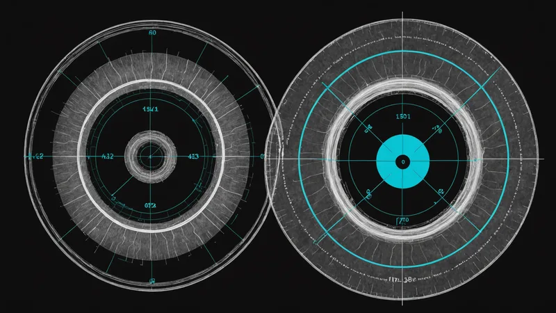
Manual nodule measurement is, despite years of experience, more variable than radiologists generally recognize. Studies of inter-reader and intra-reader measurement variability on CT nodules consistently show that the same nodule, measured by different radiologists on the same study, can differ by 1-2mm even with careful technique. The same radiologist measuring the same nodule twice will often see a 0.5-1mm difference between measurements.
This matters because the Lung-RADS category thresholds are defined in millimeters. A nodule measured at 5.8mm on the current study and documented at 5.1mm on the prior study appears to have grown 0.7mm — which is within the stable range. If a different radiologist had measured the prior at 4.9mm, the apparent growth is 0.9mm, still stable. If the measurement was 4.7mm, the apparent growth becomes 1.1mm — and whether that's significant depends on how you're applying the guideline.
This is not a negligence problem. This is the fundamental measurement precision limitation of manual nodule measurement on CT. Automated volumetric measurement, which computes lesion volume from the full three-dimensional structure rather than a single diameter, is more reproducible — it's less sensitive to the specific slice a radiologist happens to measure on, and it produces a consistent result regardless of who runs it.
Where Automated Change Detection Adds Real Value
The specific value of automated change detection in serial CT screening is not finding new nodules — that's detection, a separate capability. It's the systematic, reproducible tracking of previously documented findings across time points.
When Histolyx processes a current CT alongside the patient's prior study, it identifies the nodules documented in the prior, locates corresponding structures in the current study using deformable registration to account for respiratory phase differences and patient positioning changes, measures both the prior and current lesion dimensions in a consistent way, and flags the delta. The output isn't just the current measurements — it's the change, with confidence in the measurement approach.
This is the specific step that manual prior comparison struggles with under volume: not the initial finding, but the systematic reconciliation of current with prior. A radiologist reviewing a change detection output from Histolyx is validating the flagged changes rather than constructing the comparison from scratch. The cognitive task is confirmatory rather than generative.
We should be direct about the limitations here. Change detection based on automated registration and measurement is not error-free. Respiratory motion between studies creates registration challenges. Changes in reconstruction protocol between annual studies (different scanner, different protocol thickness) affect volumetric measurements in ways that require calibration. A 0.5mm apparent growth that falls exactly at a category threshold should be treated as equivocal regardless of whether the measurement was manual or automated. The radiologist's judgment on borderline cases remains essential — the tool reduces the frequency of borderline cases by improving measurement consistency, but it doesn't eliminate them.
The Protocol Consistency Challenge
One operational issue with serial change detection that affects imaging centers differently depending on their patient referral geography: patients who are seen at multiple imaging locations. A patient enrolled in annual CT screening at one center who moves or changes insurance may show up at a different center for their second annual study, with a prior from a different PACS system, potentially from a different scanner with a different acquisition protocol.
In this scenario, automated change detection faces a harder problem than same-protocol, same-scanner serial studies. The reconstruction kernels may differ (affecting apparent nodule density and size), the slice thickness may differ, and the DICOM metadata may format study identifiers differently. A change detection system that works well on same-site serial studies may produce less reliable change measurements on cross-site comparisons.
This isn't a problem unique to automated systems — a radiologist comparing a 1.25mm LDCT from one center to a 2.5mm standard-dose CT from another is doing a hard comparison under any workflow. But it's worth flagging because change detection outputs for cross-protocol comparisons should be interpreted with additional caution, and a well-designed system should flag protocol differences rather than presenting a measurement delta with the same confidence regardless of prior study acquisition parameters.
Integration with the Lung-RADS Reporting Framework
Lung-RADS category assignments — the 1 through 4X scale that drives follow-up recommendations — depend on both current measurement and change history. A nodule that was Lung-RADS 3 on the prior study (probably benign, 6-month follow-up recommended) may be reclassified to Lung-RADS 4A (suspicious) if it has grown more than 1.5mm in diameter, or remain Lung-RADS 3 if it's stable.
Structured reports from Histolyx include change data in a format that maps to the Lung-RADS category logic — current measurement, prior measurement, interval delta, and a computed category recommendation based on the measurement and change history. The radiologist reviews and finalizes the category, but the computational scaffolding is already in place.
For screening programs managing hundreds of patients across multi-year surveillance, the consistency this provides matters. A patient's Lung-RADS category history is the primary driver of follow-up imaging scheduling. If the category assignment is based on a consistent, reproducible measurement approach across time points rather than variable manual measurements by different radiologists, the surveillance logic is more defensible and the follow-up scheduling is more consistent.
That's the core argument for change detection: not that it's more accurate than an attentive expert radiologist on any individual case, but that at scale, across many patients over multiple years, systematic and reproducible change tracking produces better program-level surveillance quality than variable manual comparison under workload pressure.