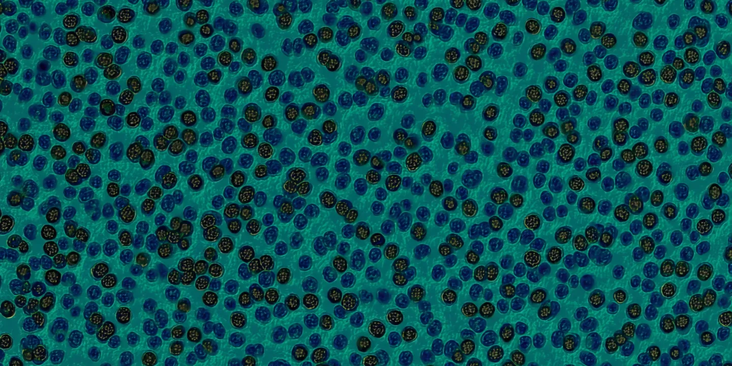
Ki-67 Scoring
Ki-67 Proliferation Index with Hotspot Detection
Global percentage positive, H-score, Allred score — plus spatial hotspot localization identifying the highest-density proliferative region.
EXAMPLE OUTPUT
Ki67_result = {
  "global_pct": 42.1,
  "hotspot_pct": 61.7,
  "h_score": 168,
  "allred_score": 6,
  "nuclei_counted": 4821,
  "hotspot_coords": [x:18240, y:9420]
}
Scoring Systems
Ki-67: Global Score vs. Hotspot — Why Both Matter
Ki-67 scoring is not simply counting brown nuclei across the whole slide. Clinical guidelines specify hotspot regions, and the choice of global vs. hotspot score changes the prognostic interpretation.
Scoring Formats Synthia Provides
Global Percentage Positive
Ki-67 positive nuclei ÷ total nuclei counted across the entire tissue area × 100. International Ki67 in Breast Cancer (IKiBC) working group recommended method for consistency.
Hotspot Percentage (Peak Region)
The highest Ki-67 proliferation index within a 400× HPF equivalent. Spatial attention maps identify the hotspot automatically — no manual high-power field selection required.
H-Score and Allred Score
Allred score (proportion + intensity, 0–8) and H-score (intensity-weighted proportion, 0–300) for centers using those historical scoring frameworks for breast cancer grading.
Clinical Context
In breast cancer, Ki-67 ≥ 20–25% is commonly used as a threshold for high proliferative activity (luminal B vs. luminal A distinction). In mantle cell lymphoma, Ki-67 > 30% defines high-risk disease. These thresholds vary by institution and guideline version.
Synthia provides the score; the pathologist interprets it in clinical context. The system deliberately outputs all three formats — global, hotspot, and H-score — so the pathologist can apply whichever framework their institution uses.
Performance Metrics
Ki-67 Internal Validation Results
Internal study, data on file. For investigational use only.
| Metric | Value | 95% CI | Reference Standard |
|---|---|---|---|
| ICC (global % positive vs. pathologist estimate) | 0.91 | 0.88–0.94 | Mean of 3 pathologist manual counts |
| ICC (H-score) | 0.93 | 0.91–0.95 | Pathologist H-score |
| Categorical concordance (<14% / 14–20% / >20%) | 89.4% | 85.4–92.6% | 3-reader majority vote |
| Hotspot localization concordance | 86.8% | 82.0–90.8% | Pathologist-identified hotspot HPF region |
| Nucleus detection recall | 94.2% | 92.5–95.7% | Manual nucleus annotation (n=5,000 cells) |
| Test-retest ICC (n=50) | 0.996 | 0.994–0.998 | Same slide, 7-day interval |
Evaluate Ki-67 Hotspot Detection on Your Cases
Request a pilot with breast carcinoma or lymphoma WSI sets. Receive global + hotspot scores with spatial heatmap overlays.